Definition
X rays are electromagnetic radiation that differentially
penetrates structures within the body and creates images of these structures on
photographic film or a fluorescent screen. These images are called diagnostic x
rays.
Purpose
Diagnostic x rays are useful in detecting abnormalities
within the body. They are a painless, non-invasive way to help diagnose
problems such as broken bones, tumors, dental decay, and the presence of
foreign bodies.
Description
X rays are a form of radiation similar to light rays,
except that they are more energetic than light rays and are invisible to the
human eye. They are created when an electric current is passed through a vacuum
tube. X rays were accidentally discovered in 1895 by German physicist Wilhem
Roentgen (1845-1923), who was later awarded the first Nobel Prize in physics
for his discovery. Roentgen was also a photographer and almost immediately
realized that the shadows created when x rays passed through the body could be
permanently recorded on photographic plates. His first x-ray picture was of his
wife's hand. Within a few years, x rays became a valued diagnostic tool of
physicians world-wide.
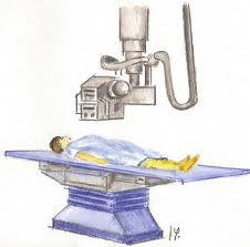
The first radiography
How x rays work
X rays pass easily through air and soft tissue of the body.
When they encounter more dense material, such as a tumor, bone, or a metal
fragment, they are stopped. Diagnostic x rays are performed by positioning the
part of the body to be examined between a focused beam of x rays and a plate
containing film. This process is painless. The greater the density of the
material that the x rays pass through, the more rays are absorbed. Thus bone
absorbs more x rays than muscle or fat, and tumors may absorb more x rays than
surrounding tissue. The x rays that pass through the body strike the
photographic plate and interact with silver molecules on the surface of the
film.
Once the film plates have been processed, dense material
such as bone shows up as white, while softer tissue shows up as shades of gray,
and airspaces look black. A radiologist, who is a physician trained to
interpret diagnostic x rays, examines the pictures and reports to the doctor
who ordered the tests. Plain film x rays normally take only a few minutes to
perform and can be done in a hospital, radiological center, clinic, doctor's or
dentist's office, or at bedside with a portable x-ray machine.
Although unnecessary exposure to radiation should be
avoided, the low levels of radiation one is exposed to during an x ray does not
cause harm with a few exceptions. Pregnant women should not have x rays unless
in emergencies the benefits highly outweigh the risks. Exposure of the fetus to
x rays, especially during early pregnancy can increase the risk of the child
later developing leukemia. Body parts not being x rayed should be shielded with
a lead apron, especially the testes, ovaries, and thyroid.

I think that x-ray machine is one of the most important inventions, because it has contributed much in the field of medicine and technology
Conclusion:
Link of a prezi presentation about X-ray machine: http://prezi.com/xsaraqzrzege/?utm_campaign=share&utm_medium=copy
No hay comentarios:
Publicar un comentario